Wednesday, January 7, 2015


Although the two pictures above are taken with the same camera settings, the left image appears more bright. The only way to control the exposure of the dental images is by setting the camera to manual mode.

Moving away from or close to the captured subject may change the image exposure even if the camera settings did not change. Moreover, tilting the camera will modify the way the light falls on the subject being captured hence the image brightness will change accordingly.

The image above to the right has been taken from the same distance used to take the image to the left. The only difference is that there is a slight change in the way the camera has been hand held. Tilting the camera in a different way in the first capture has changed the way the light fell on the teeth and sequencially the light has been reflected differently causing the increase of brightness in the image on the left.

Photography plays an important role in modern Dentistry. The ability to take professional pictures of the dental work is crucial. In the upcoming blogs,I am going to talk more about photography and share some tips and tricks in the daily practice of cosmetic dentistry.

Tuesday, November 11, 2014

Alexandria International Dental congress

This is the first post I am sharing about my lecturing experience. The 19th AIDC 2014 Dental Congress under the name of " Technology meets Challenges" was held in Alexandria, the 2nd largest city and the main port of Egypt. The organization was great and a lot of work has been done so it was obvious. The take home lesson from the congress was the importance of selecting the lecture's title. Most of the attendees do not know all of the speakers clinical and lectural background. It was clear that the  number of attendance was primarily based on the titles of the presentations . One of the lectures was titled "Ancient Dentistry"; this title was enough to determine the number of attendees in the Auditorium.On the other side, modern catchy dental subjects were able to fire the enthusiasm of the young dental generation to fill the auditoriums. My lecture title was " Progressive Smile Design,Inman Aligner Workflow". The Auditorium was full and you can tell if the presentation was successful or not by the feedback of the attendees coming to you after the lecture asking questions and showing interest to learn more about the subject presented.    
 A big thank you to all AIDC 2014 organizers and especially to Dr Mohamad Moataz Khamis,prof of Prosthodontics at Alexandria University Faculty of Dentistry.

Pictures from up down
1- pic 1—4. The lecturing auditorium 
2- pic 5 The Congress Venue from outside 
3- pic 6  Typical Egyptian Taxi 

Wednesday, March 19, 2014

Local anesthetic solution

Root canal therapy is painful but not if the patient was anesthetized very well. Today, we have a revolution in the dental material market. Solutions are always available , but you have to look into it.

Onset from Onpharma is an anesthetic product used in root canal treatments. It works by replacing a small amount of the acidic anesthetic (PH3.5) with sodium bicarbonate (PH8.4) via a mixing pen in a standard lidocain cartridge.

Benefit: This Buffered anesthetic has PH closer to the patient physiological PH allowing for more comfortable injection and shorter latency period for onset of anesthetic.


Endolce: check the sensitivity of the pulp after anesthesia by using ENDOLCE;pulp sensitivity tester.

Keywords: Endolce ,  Onset, Onpharma

Friday, January 31, 2014

Quick tip/ Necrotic pulp treatment

Necrotic pulp treatment: 

  1. Canal irrigated with 5.25 % NaOCL for 15 minutes
  2. Dry well with paper points  
  3. apply a mixture of ciprofloxacin ,metronidazole and minocycline paste as described by Hoshino et al(creamy consistency and spin down the canal with lentulo to depth of 8mm)
  4. recall,remove  preparation,irrigate,dry and obturate

Monday, December 16, 2013

There is an immense need to change the way the public views dental work.We as dentists must take to our responsibilities the task of  transforming the way the public experience dental treatments.
Luxury in Dentistry.More people are going beyond "traditional dentistry".A patient is catching up with her favorite  TV program while going through teeth whitening.No pain, no drilling , 
and instant results.Pamper yourself at Dental SPA and get a white teeth for this summer

Dr RAMI CHAYAH DMD


Thursday, October 24, 2013

Teeth whitening

There are 2 methods to choose from

1) In Office: It is a light enhanced technique done in clinic. It saves lots of time for the patient since the result is instant. Excellent option for busy people who want the result same day.

2) At home: The patient put the whitening gel himself at home in a pre- fabricated customized trays.It takes up to 21 days to get the final result.

Before doing any of them ,Dentist must check your teeth to observe any decays,stains and exposed dentin. This determines if there is potential sensitivity,treatment duration and expected result.

The procedure for:
1) In office: A whitening gel is applied to your teeth .Photos  of before situation are taken to compare the effectiveness after the treatment. Gum and lips are protected  and a coat of hydrogen peroxide is applied on teeth for 20 minutes to be removed for another coat since hydrogen peroxide gets deactivated in max 30 mins. Up to 4 coats can be applied depending on each situation.
2) At home: Two whitening trays are delivered to the patient for use at home. A whitening gel is given to be applied home.The whitening gel  can be applied for 30 minutes  every night before bedtime up to 21 days.

Now to get the best result, I use my own protocol in my practice. Most of the time the two techniques are recommended. I always ask the patient to keep the at home trays for later touch ups(maintenance). Touch ups differ from case to case.It could be every 6 months or every one year. It depends on each patient situation and life style.
Pictures are for real patient of mine

Disclaimer:This procedure should be done under the observation of a specialist. Don't try to do the above mentioned procedure at home alone. I take no responsibility for any misuse or misunderstanding. Always contact your dentist for any medical use or advise.

Dr RAMI CHAYAH DMD
Contact info: dental.spa@hotmail.com
                       tel 00961 7 754991

Thursday, January 24, 2013

what are minimal invasive burs?

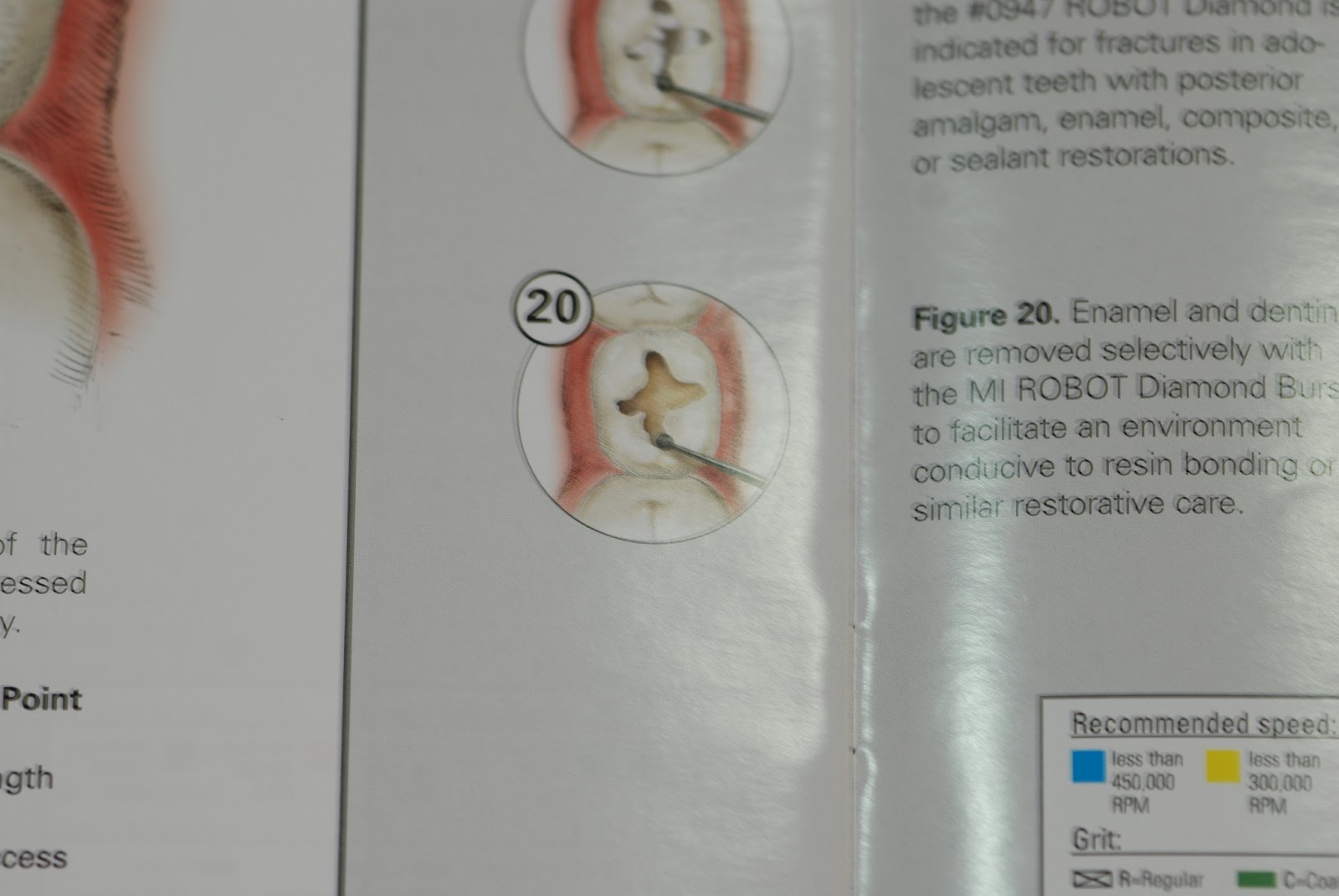
To give you a glimpse about minimal invasive burs, I will first explain the concept. Nowadays, tooth preparation techniques are totally different. In the presence of bonding materials,no need anymore to prepare tooth tissue according to Black classification.
Minimal invasive dentistry is gentle and conservative when it comes to removing carious lesions. You only remove soft pigmented dentin that does not sound. In class one,you prep teeth following carious fissures and pits only.You use the smallest bur available that can remove that carious spot and still respect anatomy.In figure 1, you see a set of burs used for minimal invasive preparations in my practice when treating carious lesions.
                                                                   Figure 1


Companies have shifted towards being  conservative by producing a whole line of burs dedicated towards minimal invasive tooth preparation. SHOFU for example included a whole section of minimal invasive burs with clear demonstrative illustrations Figure 2,3,4,5&6


                                                                        Figure 2
                                                                 Figure 3
                                                                         Figure 4
                                                                    Figure 5
                                                                         Figure 6

Finishing or just beveling the prepared sites, you can use a set of fine small finishing burs specifically selected to  just round any sharp edges left after preparation.These same burs can be used for finishing the composite after placement.In Figure 7, 8&9 you can see a set of finishing burs from German company Schezer used in my practice for this purpose.

                                                                              Figure 7
                                                                       Figure 8

                                                                   Figure 9


Sunday, October 7, 2012

Previously I blogged about oral sedation and talked about some medications. In this blog however, I will talk more about general benefits of oral sedation and its application in dentistry.

If dentist apply oral sedation in his office ,he can:

  1. Increase the quality of his procedures
  2. Increase the quantity of treatments
  3. Have more accepted treatment plans
  4. Decrease stress for the patient 
Patients who can benefit from oral sedation are so many however I will mention some of them ;

  • Complex big cases patients
  • Those who are allergic to Anesthesia
  • Phobic,Anxious and Fearful patients
  • Sever Gag reflex patients
  • Post operative sensitive patients
It is documented that 30 to 50% of patients avoid the dentist due to fear.


Spectrum of sedation has 3 phases

  1. Minimal sedation
  2. Moderate sedation
  3. Deep sedation
Dentists should sedate patients in their office within the minimal and moderate levels only. Deep sedation attempts should be avoided unless the dentist has gone
through special residency training and got special licence.

Let us mention some of the most popular oral sedatives dentists can use:

Generic                       Trade
Diazepam                     Valium
Zalepon                         Sonata
Triazolam                      Halcion
Hydroxyzine                   Atarax or Vistaril
Lorazepam                      Ativan
Nitrous Oxide                 Laughing gas


In every dental office using oral sedatives, Romazicon (flumazenil) 10ml injection 1mg/10ml or 0.1mg/ml should be available to reverse the effect of bezos in case of over sedation(overdose).

Protocols: Usually pill can be taken one to two hours before procedure. If pill will be taken home, somebody should accompany the patient and he should be informed.
A dose at night can be given as well one day before the appointment depending on the case.

Valium(Diazepam) is usually given at bedtime . Dose depends on some factors like age, BMI, history and other medications.

  • If age >or = to 65,then the dose ranges from 0 to 2.5mg 
  • If BMI >or= to 30,then the dose ranges from 5 to 10mg
The more obese the patient is the higher the dose that can be given

Patients taking CNS depressants or other serious medications should be treated carefully. You should avoid prescribing Valium to all patients with the same dosage. Please seek  consultation from experts before prescribing any medicine
in critical medical cases.

If you use oral sedatives in your office, it is recommended to track and monitor vital sign during the whole treatment appointment.Pulse oximeter blood pressure unite is necessary in this regard.  


Tuesday, September 4, 2012

T-scan computerized occlusal analysis system


T-Scan® III

computerized occlusal analysis

The only clinical diagnostic device available that senses and analyzes occlusal contact forces to quantify whether a patient's bite is balanced

Dentistry is in the right digital direction


New Version of Logicon Caries Detector Software

Carestream Dental announces the availability of Logicon Caries Detector Software version 5.0.22.4. Logicon Software is a computer-aided detection tool that helps dentists identify and treat more interproximal caries at an early stage for improved patient care.
New for Logicon Software is the addition of a PreScan feature that further automates caries detection and improves practice efficiency. With a single click, practitioners can use the PreScan feature to automatically scan all interproximal surfaces in a bitewing radiograph and immediately display the results, instead of having to scan one interproximal surface at a time. Dentists then have access to an index of the results that correlate to each interproximal surface.


Thursday, June 14, 2012

Where are we now regrading the treatment of periodontitis?

Through the past decades, it was common to treat periodontitis,periodontal pockets and bone loss, through the use of two modalities:
1) scaling and root planning
2) flaps and osseous remodeling


It was evident that the upper two modalities were unpredictable regrading the regenration of bone,fibers and the attachment around the teeth. Although scaling and root planning eliminate the bacteria from around the teeth, the long epithelium tissue would grow in around the tooth before the bone can make its regeneration,thus preventing the bone and surrounding tissue from rebuilding back around the teeth.


Nowadys, a third modality has been introduced to the bouquet;the LANAP


The LANAP (Laser Assisted New Attachment Procedure) allows removal of the inflamed epithelium,cleaves bond between calculus and root surface and kills biofilm.Thus scaling and root planning becomes effective. The final pass with the LANAP laser stimulates osseous regenration and leads to the formation of a fibronectin clot.

According to John chrispens DDS,the Laser for use in the LANAP procedure is the Nd:YAG and namely the periolase MVP-7.2.
The patient is asked not to brush in the area for one week post LANAP therapy.
Each laser works within a specific wavelength. Nd:YAG absorbes within the  range of heme(darker colors),therefore penetrates into the tissue. 
The Diode laser uses heat,therefore burns the tissue and penetrates only a short distance into the gingival sulcus before causing damage to the tissues. The CO2 and Er:YAG are absorbed by H2O, which is at the surface of oral tissues and do not penetrate into the gingival sulcus. Therfore, they have little effect on the biofilm in areas greater than 2mm subgingivally. The goal of the LANAP is not biofilm control but regenration.


Some periodontists contradict this formula. They suggest a membrane to stop epithelial cells from growing down into the socket and give bone and fibroblasts a chance to grow up.They say the laser will de-epithelialize  the pocket but the epithelium will still grow back.


The LANAP protocol:
A) Perio probe indicates excesssive pocket depth
B) Laser radiation vaporizes bacteria, diseased tissue, pathologic proteins, and alerts the practioner to the presence of tartar.
C) Ultrasonic scaler and special hand instruments are  used to remove root surface accretions.
D) Laser is used to form a gel-clot containing stem cells from bone and Periodontal ligaments.
E) Reattachment of rete ridges to clean root surface, with a stable fibrin clot at the gingival crest to create a closed system.
F) occlusal trauma adjusted
G) New attachment is regenerated


Friday, June 1, 2012

Please welcome with me MR. Opalescence PF....

I use Pola Day 9.5 % of Hydrogen Peroxide from SDI for my home whitening protocol. Today I am adding opalescence PF 35% of carbamide peroxide to the game. Opalescence PF contains potassium Nitrate, a desensitizing agent that has shown to decrease sensitivity to its minimal range.It acts more like an analgesic or anesthetic by keeping the nerve from re-polarizing after it has depolarized into the pain cycle.  This whitening system also contains fluoride that primarily acts as a tubular blocker, plugging the holes in dentin and slowing down the fluid flow that causes the sensitivity. 

Please welcome with me MR. Opalescence PF....

Please welcome with me Mr. Opalescence PF........ 

Wednesday, May 16, 2012

Periodontal disease & the heart disease; what is the link?

Periodontal disease is a risk factor for heart disease. Periodontal pathogens found in periodontitis have been found in the endothelial lining of patients with heart disease in carotid artery surgery. It was also found that DNA from periodontal pathogens have been found in cardiac tissue.
During inflammatory process,Patients predisposed to produce inflammatory mediators such as C-reactive protein, tumor necrosis factor and interleukins when the body is challenged in some way. Sometimes a patient genetically predisposed to a certain inflammatory reaction. For example in periodontal disease one may respond with high levels of C-reactive protein, which are the same inflammatory mediators seen in cardiovascular disease.
In conclusion , up to this date we can say that periodontal disease is a risk factor for heart disease but not necessarily a causing factor to this illness. More researches have to be done.

Monday, April 30, 2012

Super white teeth, here how you get it.............


 Office chairside tooth whitening

  • If you are a smoker, do not smoke for 48 hours.
  • Stay away from highly colored foods (red sauce, blueberries, etc.) and beverages (coffee, tea, etc.) for 48 hours.
  • Mild sensitivity to hot or cold liquids may occur.  This usually passes within 1-2 days.  If sensitivity is severe or persists, contact us.
  • If you experience gum sensitivity do not brush the afflicted area while brushing your teeth.
  • Immediate whitening results can be quite dramatic due to minor dehydration of your teeth.  It is normal for the color to tone down somewhat after treatment when your teeth rehydrate to a natural white tone.
  • Long term results vary from patient to patient.  This can depend on the original shade of your teeth and include habits such as smoking or drinking colored beverages (red wine, coffee, tea, etc.)
  • “Touch-up” treatments may be needed every 6-12 months to retain color.  You can ask us about different “touch-up” options such as home whitening protocol.
  • Existing fillings, crowns, bonding, etc. will not whiten.  Therefore, these may need to be changed in order to match your new smile.
  • If your family or friends notice your new smile, let them know that  whitening can help improve their smile too!
If you have any question?
email: dental.spa@hotmail.com


Follow me on twitter !

http://twitter.com/chayahdentalspa


Follow me on facebook !

http://facebook.com/dental.spa1

Saturday, April 21, 2012

Fusion presenting as gemination;rare case report

Gemination is a developmental anomaly of form ,which is recognized as an attempt by single tooth germ to divide resulting in a large tooth with a bifid crown &usually a common root and root canal in which the teeth count is normal when the anomalous tooth is counted.
Fusion is recognized as union of two separate tooth buds during odontogenesis,therefore one fewer tooth than normal in the dentition if the affected tooth is counted as one.

Case report: An 11 years old boy appeared in my clinic for examination. During intra oral diagnostics, tooth # 8 was not erupted yet. Clinically, it was revealed an abnormal maxillary temporary incisor retained in the mouth;two crowns joined togther. A provisional diagnosis  of gemination was given since the teeth count is normal. Radiographically, two fused  crowns with seperate roots and root canals were seen,along with a supernumerary tooth preventing the central incisor from erupting. A diagnosis of two temporary anomalous teeth fused together conjoined with a peg shaped anomalous supernumerary tooth embedded between the central permanent incisor and the temporary milk tooth.


A decision was made to surgically  extract the temporary tooth along  with the impacted peg shaped supernumerary tooth to give a way to the central permanent incisor to erupt.

**N.B. Normally,Maxillary central incisors begin to erupt at the age of 6. In this case the eruption of maxillary permanent right central incisor was delayed.


Friday, March 30, 2012

Chewing Gum, Aye or Nay..!


Caries is the most common chronic infectious disease in the World. We should do everything to prevent or at least decrease its spread. Sugar increases blood pressure,bad cholesterol and risk of heart disease. On top of that, sugary food adhere to teeth and stimulate plaque growth.
Xylitol is a five carbon sugar alcohol compound. Sugar alcohols are neither sugar nor alcohol. Xylitol is not an artificial sweetener as well. Xylitol is a naturally sugar compound which can be derived from  vegetables and plants. Xylitol has all the benefits of sugar without its many drawbacks.
Bacteria can use the six carbon monosachcharide such as glucose or fructose as food source to produce plaque on teeth which leads to decay but cant use xylitol for this same purpose.

Xylitol has been added to chewing gums as a sweetener instead of sugar.For this reason, it is very important to know all its advantages. Now to get back to our main subject about chewing gums. Chewing gums are very effective in preventing teeth decay for the following reasons:
  1. It increases saliva flow up to 10 times.Saliva helps to wash away food debris and plaque.
  2. Chewing gum mechanically washes away food debris and food trapped on deep areas of back teeth.
  3. The increased saliva flow increases remineralization process. What the plaque acids take out, the extra saliva puts back.
  4. Dry mouth is common for people who take medications such as antidepressants and anti-histamines. More saliva production helps those patients from dry mouth side effect.
  5. Chewing gum  improves memory and increases concentration. We all remember the legend Micheal Jordan and how he used to chew gum during his NBA games. 
  6. Chewing gum helps those who want to quit smoking. 

How to use the chewing gum?

Chew gum immediately after each meal for 20 minutes.When you eat sugary meals, the PH level in your mouth drops rapidly and remains low for some time.The chewing action stimulates far more saliva,which rapidly brings the PH levels back up to its normal limit.

N.B. Be sure to chew  chewing gum with xylitol.Other sweeteners such as mannitol,aspartame or sorbitol can be used by bacteria in the mouth as food sources and they can lead to causing decay. So be sure whenever you buy any product with sugar substitute,that xylitol is the main and only ingredient.

Saturday, March 24, 2012

Fluoride treatment

Fluoride treatment can reduce caries in children and adults.
There are many Types of fluoride treatments, we will take about topical method.
Topical fluoride application includes either sodium fluoride NaF or Acidulated phosphate fluoride APF.
Sodium fluoride comes in a solution, gel or foam forms of 2 % . It is also available in varnish of 5 % . Acidulated phosphate fluoride comes in a solution, gel or foam of 1.23%.
APF is the preferred topical application over NaF, however it is avoided when patient has cosmetic restorations as it may cause some pitting.

Fluoride application techniques:
1- paint-on-method: not for gel or foam forms to avoid fluoride ingestion.
2- tray technique for gel or foam forms; 2 ml for children & 5 ml for adults.
In both techniques, dry teeth before & after with 2*2 gauze.
No drinking or eating for 30 min after procedure.

3- fluoride varnish application: safe and durable. Most effective technique to use on children.

Optimal level of fluoride in drinking water is 0.7mg/ L .

Ingestion of extra fluoride is toxic.
Certainly lethal dose in adults is 5 to 10 grams of sodium fluoride taken at one time. It is between 0.5-1g in children depending on size and weight.
Signs of toxicity begin to appear after 30 min of ingestion.
Emergency treatment : induce vomiting by stimulating back of throat or by Ipecac syrup.
Giving calcium binding agent such as milk.

Friday, March 23, 2012

Drying root canals

While I was drying root canal yesterday for tooth number 4, I noticed a very important thing. Regardless of how copious you try to irrigate the canal with disinfectant, small tissue tags can still resist the force of irrigation.
Very important tip: always while you are drying the canal with paper points at your last step, put three or four paper points together into the canal until they fit tight, then twist them all together against the canal walls. This way, you mechanically remove any tiny small tag holding still on the inner walls.If you find any tags trapped on the paper points, repeat irrigation and drying, before filling the canal with GP.

Friday, March 16, 2012

Sutures -everything you should know

Suture material that retains long term strength for up to 14 days and resorbs in 21 to 28 days is polyglycolic acid (PGA )

Conversely suture to be placed intraorally where tissue heals quickly ; surgical gut or the new rapidly resorbable PGA suture material (PGA -FA) is used.


Degradation of absorbable , biological origin sutures such as surgical gut ( plain & chromic gut) are digested by intra oral enzymes .

Gut suture is from animal protein
Which resorbs in 1 to 2 days. If coated with chromic compound, it extends 7 to 10 days.

Fast absorbing PGA FA suture is made from synthetic polymers and dissolves in 7 to 10 days. It should be used when gut suture is contraindicated .

Thread sutures range in diameter from 0 to 10

The higher the number, the thinner and more delicate the thread.


5-0 diameter is used in plastic perio surgery
4- 0 diameter is used for other perio flaps.

3-0 diameter used in implant surgeries when mattress suturing technique is used. 4-0 is used when interrupted or continuos techniques are used.


Silk is a classical suture used in dentistry . It's nonresorbable inexpensive multifilament material. Dentist should remove it in a week. It's down side is pulling bacteria into the wound site. In this case nonresorbable sutures such as nylon, polyester, polyethylene, polypropylene or expanded polytetrafluoroethylene (e-PTFE) are a better choice.


Polyester sutures are made of multifilaments braided in a single strand. This suture is coated with absorbable compound that makes the knot untie if not secured well by the dentist

e-PTFE is better in this regard . It is nonresorbable monofilament that has high tensile strength but a little more expensive than polyester.

The 3/8and 1/2 are the most used suture needles in dentistry .


The 3/8 passes easily from buccal to lingual in one motion

The 1/2 circle needle is used in more restricted areas such as maxillary molar area and anterior incisal areas.


Generally in dentistry the 3/8 reverse cutting needle with 3-0 or 4-0 thread diameter and the 1/2 reverse cutting needle with the thinner more delicate 5-0 or 6-0 thread diameter are most commonly used.